Overview
The diSPIM is a flexible and easy-to-use implementation of Selective Plane Illumination Microscopy (SPIM) that allows for dual views (d) of the sample while mounted on an inverted (i) microscope.
The diSPIM has two (usually symmetric) optical paths for light sheet imaging. Two objectives are placed at right angles above a sample mounted horizontally. A light sheet is created from one objective and then imaged using the other objective. A stack of images is collected by moving the light sheet through the sample. The role of the two objectives can be exchanged to collect another stack from a perpendicular direction, and then the two datasets can be computationally merged to yield a 3D dataset with isotropic resolution.
The diSPIM “head” can be mounted on various inverted microscopes. A microscope-specific adapter bracket accepts the SPIM-MOUNT to which the SPIM assembly attaches.1) Thus far brackets exist for the following microscopes:
- ASI RAMM frame (shown in figure below)
- Leica DMI-6000
- Nikon TE-300, Ti
- Olympus IX-71/81, IX-73/83
- Zeiss Axio-Observer
The rest of this document will concentrate on the function, alignment and operation of the diSPIM microscope. The vast majority of the instructions apply regardless of the inverted microscope frame on which the diSPIM “head” is mounted.
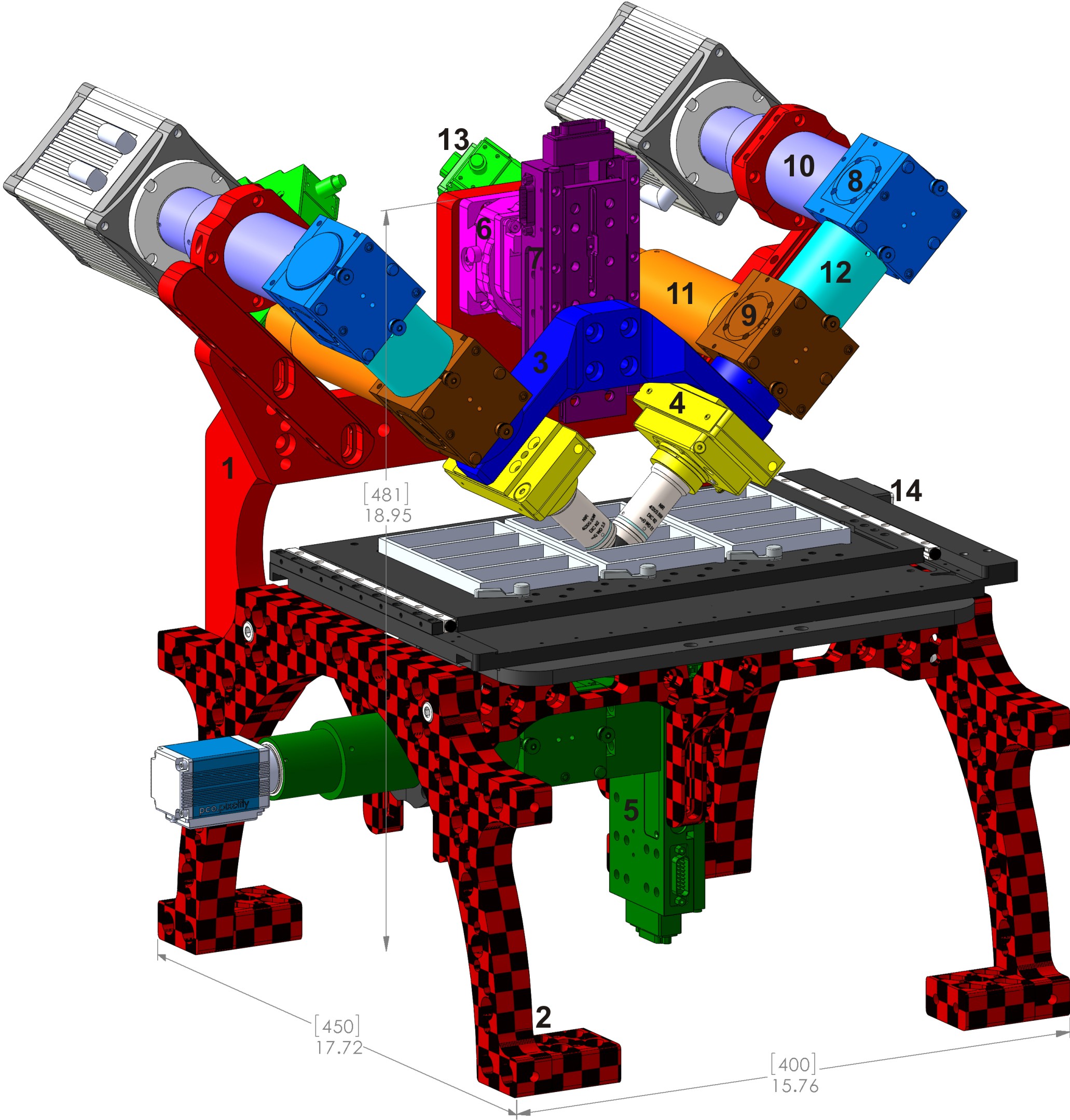
Figure 1 shows the main components of the diSPIM as mounted on a RAMM frame.
 Figure 1: Typical complete diSPIM system on RAMM frame showing major components. 1) SPIM-MOUNT , 2) SPIM-RAMM , 3) SPIM arm mount (RAO-0046 ), 4) piezo objective mover APZOBJ-200 or similar, 5) MIM inverted microscope with LS-50M stage, 6) CDZ-1000 centering stage or CDZ-R block, 7) LS-50M stage, 8) MIM-CUBE-II w/ mirror, 9) MIM-CUBE-II w/ fluorescence filters, 10) C60-TUBE_B imaging tube lens, 11) C60-TUBE_160 scanner tube lens, 12) 50mm extension tube, 13) MM-SCAN_1M light sheet scanner, 14) MS-2500 XY stage.